| Record Information |
|---|
| Version | 1.0 |
|---|
| Creation Date | 2016-05-25 18:09:04 UTC |
|---|
| Update Date | 2016-11-09 01:17:20 UTC |
|---|
| Accession Number | CHEM021646 |
|---|
| Identification |
|---|
| Common Name | Iduronic acid |
|---|
| Class | Small Molecule |
|---|
| Description | |
|---|
| Contaminant Sources | - FooDB Chemicals
- HMDB Contaminants - Urine
|
|---|
| Contaminant Type | Not Available |
|---|
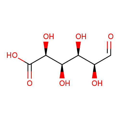
| Chemical Structure | |
|---|
| Synonyms | | Value | Source |
|---|
| Iduronate | Generator | | L-Iduronate | HMDB | | L-Iduronic acid | HMDB | | Acid, iduronic | HMDB | | (2S,3R,4R,5S)-2,3,4,5-Tetrahydroxy-6-oxohexanoate | HMDB | | Iduronic acid | MeSH |
|
|---|
| Chemical Formula | C6H10O7 |
|---|
| Average Molecular Mass | 194.139 g/mol |
|---|
| Monoisotopic Mass | 194.043 g/mol |
|---|
| CAS Registry Number | 3402-98-0 |
|---|
| IUPAC Name | (2S,3R,4R,5S)-2,3,4,5-tetrahydroxy-6-oxohexanoic acid |
|---|
| Traditional Name | iduronic acid |
|---|
| SMILES | O[C@H](C=O)[C@H](O)[C@@H](O)[C@H](O)C(O)=O |
|---|
| InChI Identifier | InChI=1S/C6H10O7/c7-1-2(8)3(9)4(10)5(11)6(12)13/h1-5,8-11H,(H,12,13)/t2-,3+,4-,5+/m1/s1 |
|---|
| InChI Key | IAJILQKETJEXLJ-LECHCGJUSA-N |
|---|
| Chemical Taxonomy |
|---|
| Description | belongs to the class of organic compounds known as glucuronic acid derivatives. Glucuronic acid derivatives are compounds containing a glucuronic acid moiety (or a derivative), which consists of a glucose moiety with the C6 carbon oxidized to a carboxylic acid. |
|---|
| Kingdom | Organic compounds |
|---|
| Super Class | Organic oxygen compounds |
|---|
| Class | Organooxygen compounds |
|---|
| Sub Class | Carbohydrates and carbohydrate conjugates |
|---|
| Direct Parent | Glucuronic acid derivatives |
|---|
| Alternative Parents | |
|---|
| Substituents | - Glucuronic acid or derivatives
- Hexose monosaccharide
- Medium-chain hydroxy acid
- Medium-chain fatty acid
- Beta-hydroxy acid
- Hydroxy fatty acid
- Monosaccharide
- Fatty acyl
- Hydroxy acid
- Fatty acid
- Alpha-hydroxy acid
- Beta-hydroxy aldehyde
- Alpha-hydroxyaldehyde
- Secondary alcohol
- Polyol
- Monocarboxylic acid or derivatives
- Carboxylic acid
- Carboxylic acid derivative
- Aldehyde
- Carbonyl group
- Alcohol
- Organic oxide
- Hydrocarbon derivative
- Aliphatic acyclic compound
|
|---|
| Molecular Framework | Aliphatic acyclic compounds |
|---|
| External Descriptors | Not Available |
|---|
| Biological Properties |
|---|
| Status | Detected and Not Quantified |
|---|
| Origin | Not Available |
|---|
| Cellular Locations | Not Available |
|---|
| Biofluid Locations | Not Available |
|---|
| Tissue Locations | Not Available |
|---|
| Pathways | Not Available |
|---|
| Applications | Not Available |
|---|
| Biological Roles | Not Available |
|---|
| Chemical Roles | Not Available |
|---|
| Physical Properties |
|---|
| State | Not Available |
|---|
| Appearance | Not Available |
|---|
| Experimental Properties | | Property | Value |
|---|
| Melting Point | Not Available | | Boiling Point | Not Available | | Solubility | Not Available |
|
|---|
| Predicted Properties | |
|---|
| Spectra |
|---|
| Spectra | | Spectrum Type | Description | Splash Key | View |
|---|
| Predicted GC-MS | Predicted GC-MS Spectrum - GC-MS (Non-derivatized) - 70eV, Positive | splash10-000i-9700000000-cdaf9f94e02e4e7ba7d2 | Spectrum | | Predicted GC-MS | Predicted GC-MS Spectrum - GC-MS (5 TMS) - 70eV, Positive | splash10-009i-4212940000-02a8091c226f3a41d70e | Spectrum | | Predicted GC-MS | Predicted GC-MS Spectrum - GC-MS (Non-derivatized) - 70eV, Positive | Not Available | Spectrum | | Predicted LC-MS/MS | Predicted LC-MS/MS Spectrum - 10V, Positive | splash10-056s-2900000000-86606b8382ffe403bc8e | Spectrum | | Predicted LC-MS/MS | Predicted LC-MS/MS Spectrum - 20V, Positive | splash10-0a4i-9500000000-af67bc97552641417bee | Spectrum | | Predicted LC-MS/MS | Predicted LC-MS/MS Spectrum - 40V, Positive | splash10-0a4i-9100000000-205a7cff1e298283a080 | Spectrum | | Predicted LC-MS/MS | Predicted LC-MS/MS Spectrum - 10V, Negative | splash10-05p9-9700000000-ee070fab487c9f3e49b7 | Spectrum | | Predicted LC-MS/MS | Predicted LC-MS/MS Spectrum - 20V, Negative | splash10-0a4r-9400000000-284825e422ba9585f17b | Spectrum | | Predicted LC-MS/MS | Predicted LC-MS/MS Spectrum - 40V, Negative | splash10-0a4i-9100000000-64d4f9d758e2a4eeba68 | Spectrum | | Predicted LC-MS/MS | Predicted LC-MS/MS Spectrum - 10V, Negative | splash10-00fr-9300000000-77aa1619935fe785e397 | Spectrum | | Predicted LC-MS/MS | Predicted LC-MS/MS Spectrum - 20V, Negative | splash10-0a6r-9000000000-da4be7e32a829503c021 | Spectrum | | Predicted LC-MS/MS | Predicted LC-MS/MS Spectrum - 40V, Negative | splash10-0a6r-9000000000-c1c9308cd925f43d015f | Spectrum | | Predicted LC-MS/MS | Predicted LC-MS/MS Spectrum - 10V, Positive | splash10-01y5-7900000000-df218a4e340a0ff668c1 | Spectrum | | Predicted LC-MS/MS | Predicted LC-MS/MS Spectrum - 20V, Positive | splash10-03kl-9100000000-d22d4d2644d77203331d | Spectrum | | Predicted LC-MS/MS | Predicted LC-MS/MS Spectrum - 40V, Positive | splash10-08fu-9000000000-93301767b758d212114f | Spectrum | | 1D NMR | 1H NMR Spectrum | Not Available | Spectrum | | 1D NMR | 13C NMR Spectrum | Not Available | Spectrum | | 1D NMR | 1H NMR Spectrum | Not Available | Spectrum | | 1D NMR | 13C NMR Spectrum | Not Available | Spectrum | | 1D NMR | 1H NMR Spectrum | Not Available | Spectrum | | 1D NMR | 13C NMR Spectrum | Not Available | Spectrum | | 1D NMR | 1H NMR Spectrum | Not Available | Spectrum | | 1D NMR | 13C NMR Spectrum | Not Available | Spectrum | | 1D NMR | 1H NMR Spectrum | Not Available | Spectrum | | 1D NMR | 13C NMR Spectrum | Not Available | Spectrum | | 1D NMR | 1H NMR Spectrum | Not Available | Spectrum | | 1D NMR | 13C NMR Spectrum | Not Available | Spectrum | | 1D NMR | 1H NMR Spectrum | Not Available | Spectrum | | 1D NMR | 13C NMR Spectrum | Not Available | Spectrum | | 1D NMR | 1H NMR Spectrum | Not Available | Spectrum | | 1D NMR | 13C NMR Spectrum | Not Available | Spectrum | | 1D NMR | 1H NMR Spectrum | Not Available | Spectrum | | 1D NMR | 13C NMR Spectrum | Not Available | Spectrum | | 1D NMR | 1H NMR Spectrum | Not Available | Spectrum | | 1D NMR | 13C NMR Spectrum | Not Available | Spectrum |
|
|---|
| Toxicity Profile |
|---|
| Route of Exposure | Not Available |
|---|
| Mechanism of Toxicity | Not Available |
|---|
| Metabolism | Not Available |
|---|
| Toxicity Values | Not Available |
|---|
| Lethal Dose | Not Available |
|---|
| Carcinogenicity (IARC Classification) | Not Available |
|---|
| Uses/Sources | Not Available |
|---|
| Minimum Risk Level | Not Available |
|---|
| Health Effects | Not Available |
|---|
| Symptoms | Not Available |
|---|
| Treatment | Not Available |
|---|
| Concentrations |
|---|
| Not Available |
|---|
| External Links |
|---|
| DrugBank ID | Not Available |
|---|
| HMDB ID | HMDB0002704 |
|---|
| FooDB ID | FDB023047 |
|---|
| Phenol Explorer ID | Not Available |
|---|
| KNApSAcK ID | Not Available |
|---|
| BiGG ID | 48353 |
|---|
| BioCyc ID | Not Available |
|---|
| METLIN ID | 3331 |
|---|
| PDB ID | Not Available |
|---|
| Wikipedia Link | Iduronic acid |
|---|
| Chemspider ID | 17794 |
|---|
| ChEBI ID | 24769 |
|---|
| PubChem Compound ID | 18845 |
|---|
| Kegg Compound ID | C06472 |
|---|
| YMDB ID | Not Available |
|---|
| ECMDB ID | Not Available |
|---|
| References |
|---|
| Synthesis Reference | Not Available |
|---|
| MSDS | Not Available |
|---|
| General References | | 1. Roden, Lennart; Dorfman, Albert. Metabolism of mucopolysaccharides in mammalian tissues. V. Origin of L-iduronic acid. Journal of Biological Chemistry (1958), 233 1030-3. | | 2. Fuller M, Meikle PJ, Hopwood JJ: Glycosaminoglycan degradation fragments in mucopolysaccharidosis I. Glycobiology. 2004 May;14(5):443-50. Epub 2004 Jan 12. | | 3. Cheng F, Petersson P, Arroyo-Yanguas Y, Westergren-Thorsson G: Differences in the uptake and nuclear localization of anti-proliferative heparan sulfate between human lung fibroblasts and human lung carcinoma cells. J Cell Biochem. 2001;83(4):597-606. | | 4. Inoue H, Otsu K, Suzuki S, Nakanishi Y: Difference between N-acetylgalactosamine 4-sulfate 6-O-sulfotransferases from human serum and squid cartilage in specificity toward the terminal and interior portion of chondroitin sulfate. J Biol Chem. 1986 Apr 5;261(10):4470-5. | | 5. Ginsberg LC, Di Ferrante DT, Di Ferrante N: A substrate for direct measurement of L-iduronic acid 2-sulfate sulfatase. Carbohydr Res. 1978 Jul;64:225-35. | | 6. Casu B, Petitou M, Provasoli M, Sinay P: Conformational flexibility: a new concept for explaining binding and biological properties of iduronic acid-containing glycosaminoglycans. Trends Biochem Sci. 1988 Jun;13(6):221-5. | | 7. Gigli M, Ghiselli G, Torri G, Naggi A, Rizzo V: A comparative study of low-density lipoprotein interaction with glycosaminoglycans. Biochim Biophys Acta. 1993 Apr 7;1167(2):211-7. | | 8. Bartolucci C, Cellai L, Iannelli MA, Lamba D, Liverani L, Mascellani G, Perola E: Inhibition of human leukocyte elastase by chemically and naturally oversulfated galactosaminoglycans. Carbohydr Res. 1995 Oct 23;276(2):401-8. | | 9. Mulloy B, Forster MJ: Conformation and dynamics of heparin and heparan sulfate. Glycobiology. 2000 Nov;10(11):1147-56. |
|
|---|